He May Look Creepy, But This ‘Manikin’ Could Help Save Your Life
The University of Hawaiʻi John A. Burns School of Medicine is using a new high-tech human patient simulator to train future doctors.
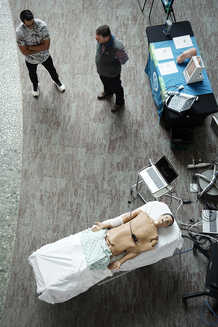
A topless patient lies motionless in the lobby. His chest rises and falls rhythmically. Medical student Ryan Keli‘i Shontell and Dr. Benjamin Berg approach the gurney. Then Berg pulls the skin covering the patient’s torso off.
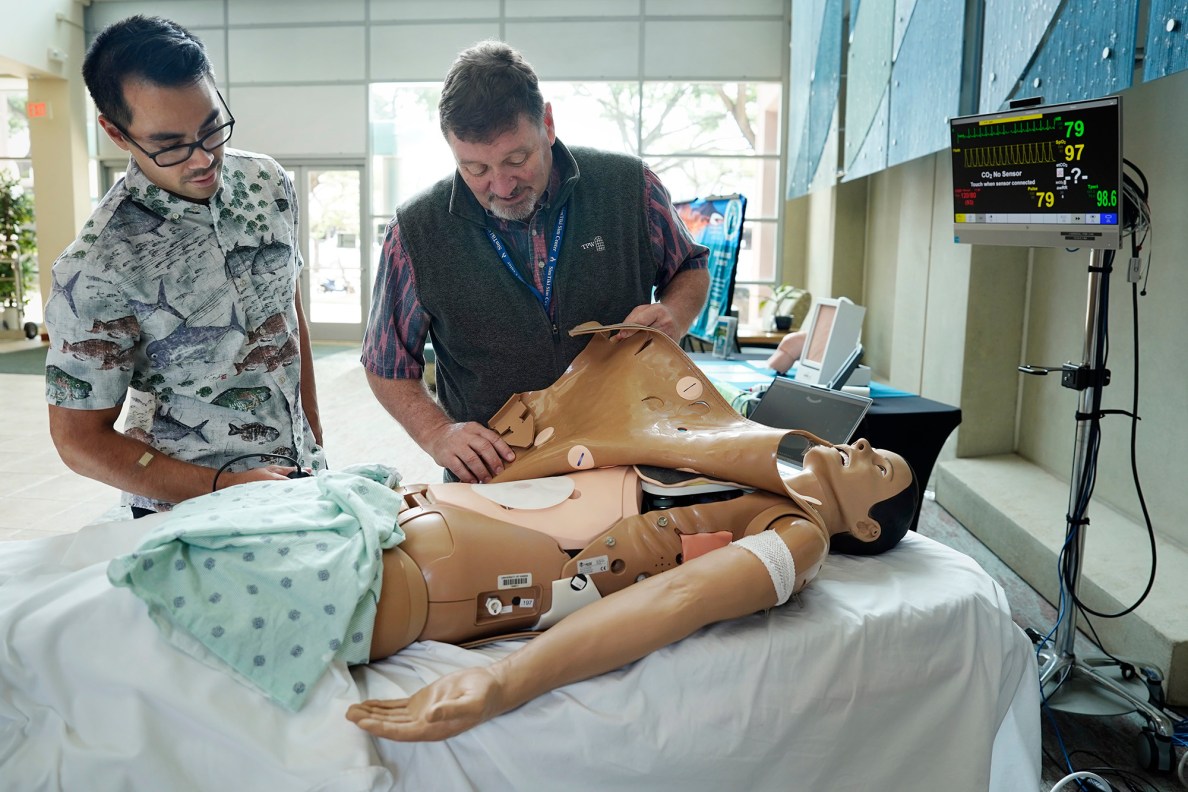
“This is the Laerdal high-fidelity human patient simulator, a critical care and anesthesia manikin,” Kris Hara, JABSOM operations director and chief simulation specialist, said. “This simulator type costs about $50,000.”
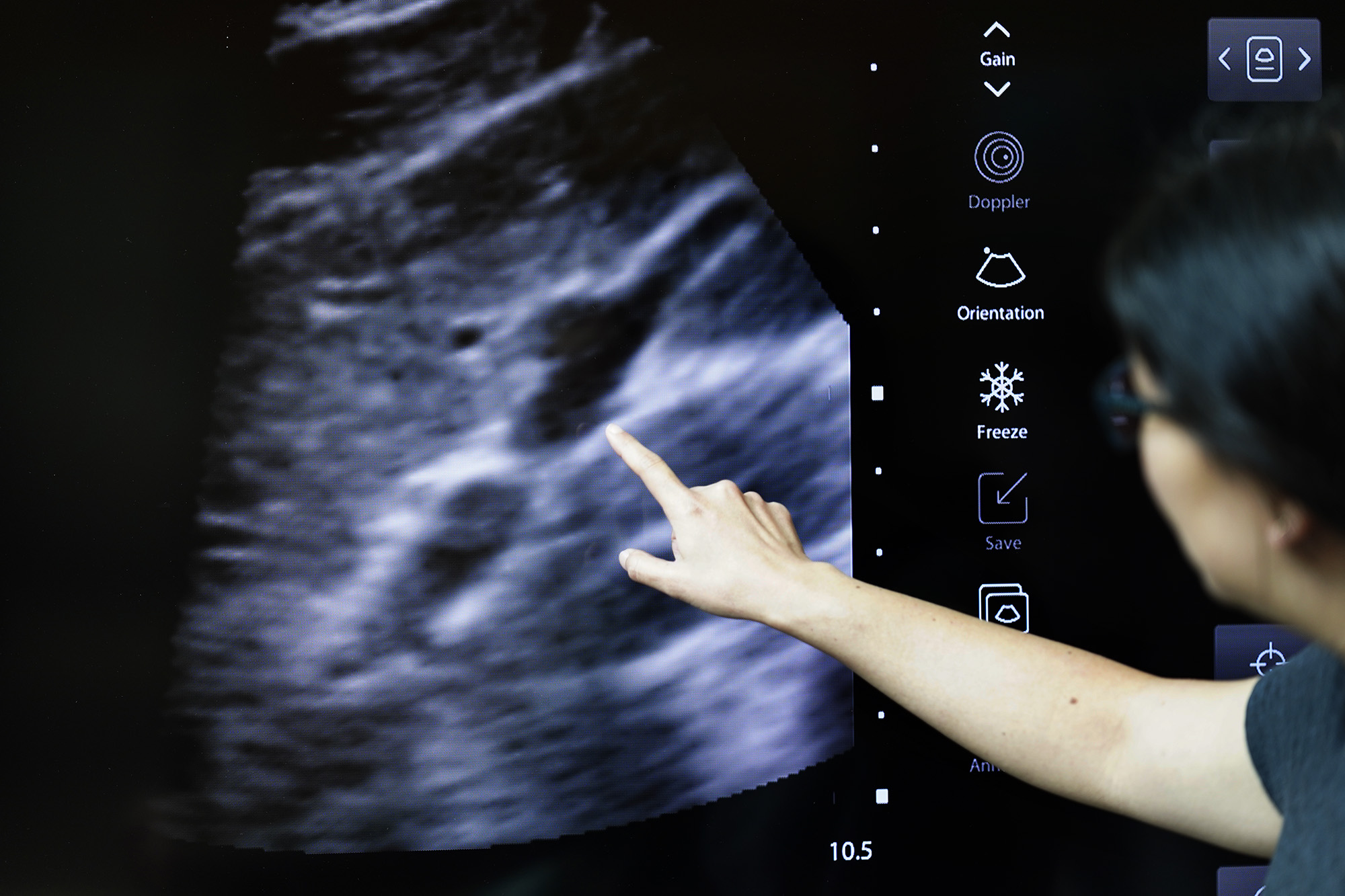
A “manikin” differentiates the human-like figure from a mannequin that models clothes. This one does more than show off the latest hospital gown. It’s filled with RFID tags — radio-frequency identification tags that use radio waves to identify objects — to teach students how to see with an ultrasound probe. “You get to see the insides of real patients,” Berg said, pointing to the monitor.

Students get hands-on training with a manikin before working with real, human patients.
“Adding ultrasound capabilities brings an important level of realism to simulations because point-of-care ultrasound has become such an important part of current medical practice,” Shontell, in his fourth year of studies at the medical school, said.
As second-year student Hollis Tam moved the ultrasound probe along the manikin’s torso, Berg guided his hand and probe toward an RFID tag. Tam said it provided a realistic learning environment for diagnostic techniques he will use in a real-world clinical scenario.
“It’s a great opportunity to practice using and reading an ultrasound,” Tam said.
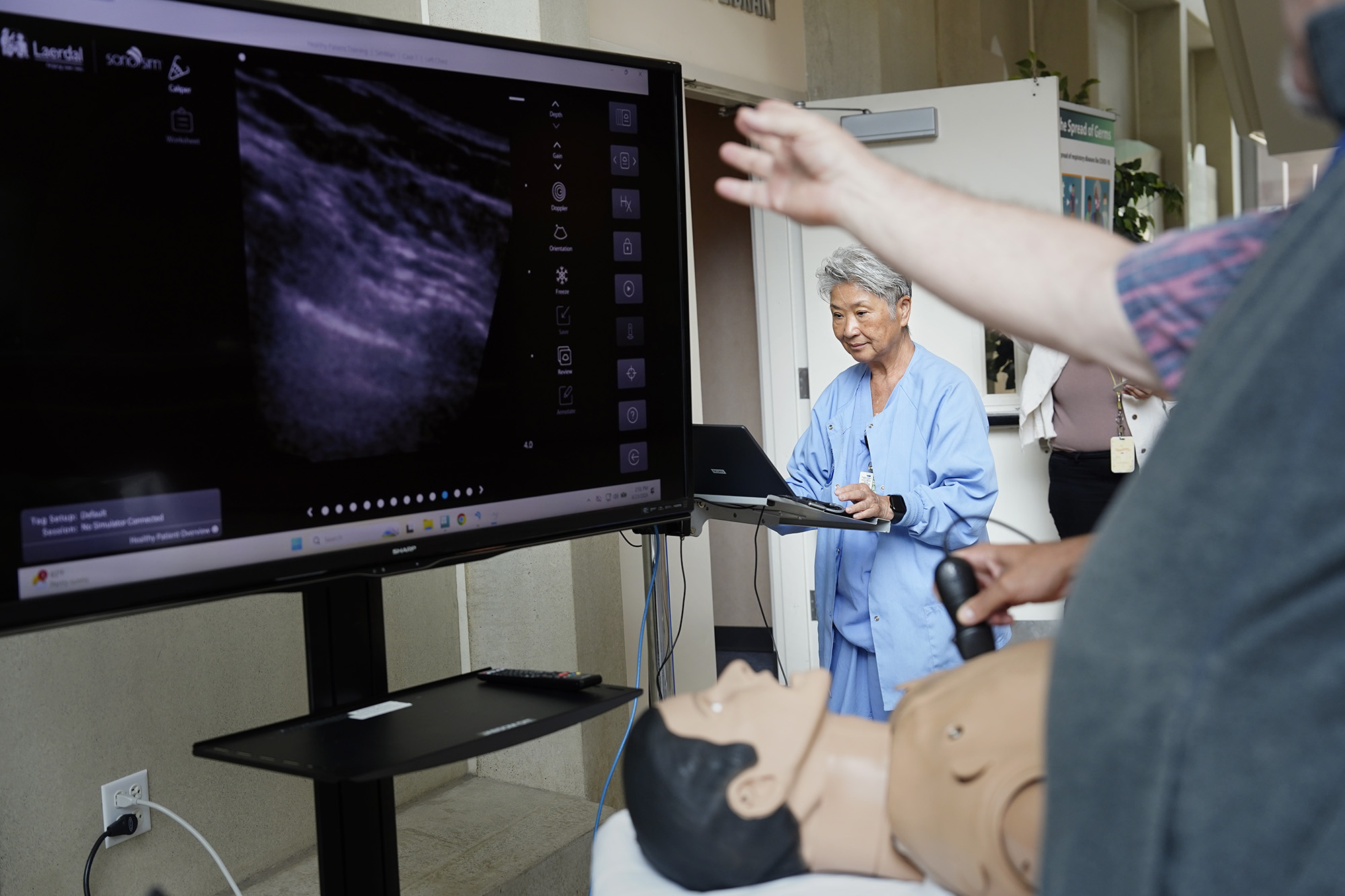
“The applications are enormous,” Shontell said. “They continue to expand as ultrasound becomes more widely incorporated into patient care.” He cited examples such as using ultrasounds in the emergency room to rapidly identify life-threatening conditions like a collapsed lung or internal bleeding. Or using them in a primary care provider’s office where they could identify fluid buildup around the lungs or abdomen.

“There’s a rib,” Berg said, while a student moved the ultrasound probe over the manikin.

During preclinical years at the medical school, Shontell said, students participate in several workshops to learn basic ultrasound techniques by practicing on each other. Because the students are typically heatlhy, however, they don’t get to see the ailments — such as a collapsed lung — that they will ultimately be looking for as doctors.
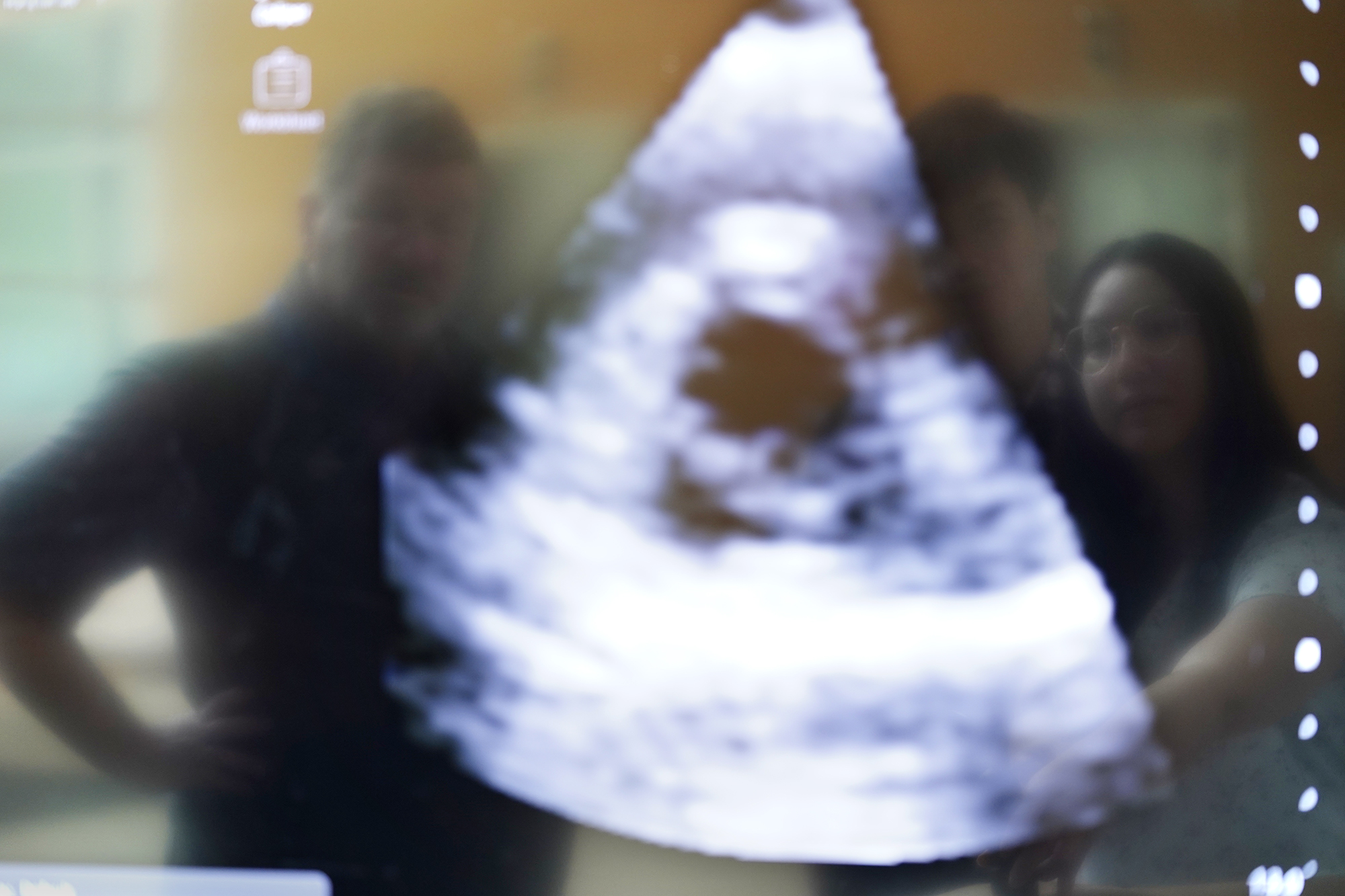
Shontell said the greatest strength of incorporating ultrasound into the SimTiki Simulation Center is the ability to simulate diseases so that students can recognize what abnormal findings actually look like. “That kind of exposure is difficult to obtain when training on healthy volunteers,” he said.
“It was exciting to try,” Shontell said, “because it offers an opportunity for future students to practice identifying important abnormalities in a low-stakes environment before encountering them in real patients.”
The manikin remained motionless as Berg wrapped up talking story with a couple of students.
“I’m feeling much better now,” the human patient simulator said. “Thank you.”